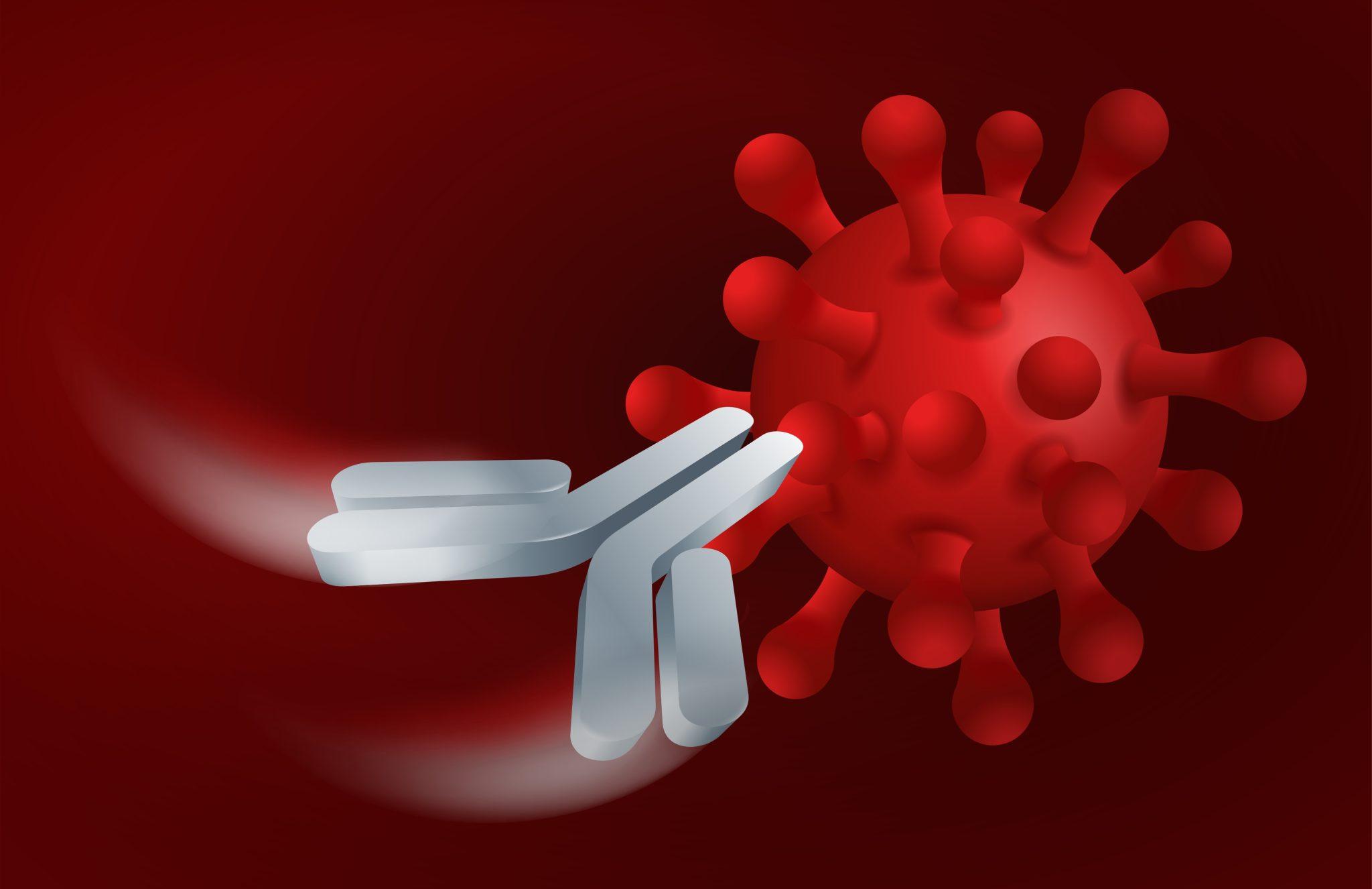
Nanobodies are the smallest functional single-domain antibodies known to be able to stably bind to antigens, and have unique structural and functional advantages. The molecular weight of nanobodies is only 12-15 kDa, which retains the antigen binding ability of traditional antibodies. However, nanobodies have higher solubility and stability, and have unique advantages in biological function and biochemical characteristics. Therefore, nanobodies have shown good control effects in disease diagnosis, cancer and infectious diseases.
What are Nanobody-Drug Conjugates (NDCs)
The development concept of nanobody-drug conjugates (NDCs) is similar to that of antibody drug conjugates (ADC), that is, the antibody in ADC is replaced by a nanobody with higher affinity. Because nanobodies are smaller in size and have stronger tumor tissue penetration capabilities, researchers are currently applying them to ADC drugs for the treatment of solid tumors, hoping to improve the solid tumor tissue penetration effects of current ADC drugs.
Nanobody Structure
Generally, the relative molecular mass of nanobodies is about 15kDa, which is only about 1/10 of traditional antibodies. In addition, the crystal structure of the nanobody is a rugby ball shape with a diameter of about 2.5nm and a length of about 4.2nm. The unique molecular structure enables it to have good tissue penetration, shorter half-life and higher renal clearance rate, and can penetrate the blood-brain barrier. Structurally, nanobodies are composed of complementarity-determining regions and framework regions. The complementarity determining regions include CDR1, CDR2 and CDR3. During the differentiation of B cells in camelid animals, the recombinant genes produced by VDJ rearrangement are involved in the coding of the CDR3 region. Therefore, the CDR3 region is relevant to the specificity of nanobodies. The length range of the nanobody CDR3 region from 3 to 28 amino acids ensures the storage capacity of the nanobody library.
Nanobodies vs Antibodies
Traditional IgG antibodies are composed of two heavy chains and two light chains. The heavy chain includes three constant domains: CH1, CH2 and CH3, and a variable domain VH. The light chain of an IgG antibody contains a variable domain VL and a constant domain CL. IgG-derived antibody drugs include IgG antibodies, bispecific antibodies, Fab fragment antibodies and scFv fragment antibodies.
Compared with the CDR3 region of traditional antibodies, which only has 8 to 15 amino acids, the longer CDR3 region of some nanobodies is expected to help them recognize hidden epitopes of antigens. The backbone region includes FR1, FR2, FR3 and FR4. There are four hydrophilic amino acid mutations on FR2, which improves water solubility. The special disulfide bond between CDR1 and CDR3 enhances the stability of antibodies under conditions such as high pressure, high temperature, and denaturants, which is beneficial to the production and preservation of nanobodies and also creates the possibility of new drug delivery methods. Nanobodies have numerous potential advantages over mAbs or their fragments in drug development:
Tissue penetration: The larger size of mAbs and the presence of the Fc region can improve pharmacokinetic properties, but also affects their ability to penetrate tissues. Smaller volume Nbs can achieve higher tissue penetration and stronger cell killing in vivo.
Antigen recognition ability: Among all complementarity determining regions, CDR3 accounts for 60-80% of the antigen recognition specificity. The CDR3 loop is long and extended. This characteristic gives VHH better antigen recognition specificity and affinity, which can enhance the recognition of hidden tumor antigen epitopes.
Strong stability: Compared with mAbs, Nbs have significantly high thermal stability, Tm value and reversible thermal denaturation. In addition, Nbs can resist the denaturing effects of proteases, extreme pH, and hydrophobic agents.
Concatenated into multi-specific or multivalent configurations: Compared with mAbs, Nbs are smaller in size, which is conducive to tandem with a variety of antibody domains with different functions for the delivery of multi-functional drugs, enhancing affinity while avoiding rapid renal clearance and increasing drug ratio.
Nanobody Preparation Process
Step 1: Design antigen to immunize camel-derived animals, that is, design a hapten based on the structure of a macromolecule or a small molecule, and couple the protein to make an antigen to immunize camel-derived animals. Generally, 4 to 5 times of immunization are required.
Step 2: Extract RNA library for screening. That is, blood is first taken, RNA from lymphocytes in the blood is extracted, reverse transcribed into cDNA, and then PCR amplification is performed. After electroporation into competent cells, a phage library is established, and multiple positive clones are obtained through phage display technology.
Step 3: Expression and purification of positive clones. Use the icELISA detection method to test the plate to determine its antigen-binding activity and inhibition rate, then store the bacteria and express and purify the existing nanobody active protein in large quantities. Finally, corresponding detection methods were established to evaluate the antigen-binding specificity, stability and other aspects of the obtained nanobodies and applied them.
Advantages of Nanobodies
High Stability
Unlike traditional antibodies, nanobodies can be refolded after chemical and thermal denaturation, and disulfide bonds are re-formed between the complementary determining region 1 (CDR1) and the complementary determining region 3 (CDR3), maintaining antibody activity and regaining antigen binding ability, thus showing higher stability. The study found that nanobodies can still maintain more than 80% of their biological activity after being placed at 37°C for 1 week, which makes nanobodies easier to use and store at room temperature. In addition, traditional antibodies and other types of genetically engineered antibodies will fail or break down under denaturing conditions of high heat, surfactants, proteases and extreme pH, while nanobodies remain highly stable. This allows the nanobodies to resist the effects of strong acids and proteases in the stomach and intestines, prolong the action time in the body, and enable disease diagnosis and treatment.
High Specificity and Affinity
The antigen-binding surfaces of traditional antibody Fab fragments and single-chain antibody scFv often form a concave topology, and usually can only recognize sites located on the antigen surface. Nanobodies are small in size and their long CDR3 can form a stable convex ring structure. In addition, the smaller size of nanobodies is conducive to the reduction of steric hindrance, so nanobodies can penetrate deep into the antigen to better recognize and bind to antigenic epitopes that traditional antibodies cannot recognize, thereby improving antigen specificity and affinity. In addition, some studies have found that the cysteine contained in the convex ring of the CDR3 nanobody can stabilize the convex ring structure of CDR3 by forming a disulfide bond with the CDR1 ring, thereby improving the stability and binding ability of the nanobody.
Strong Tissue Penetration and Weak Immunogenicity
Compared with traditional antibodies, the smaller size of nanobodies makes it easier to penetrate various tissues of the body and can effectively penetrate the blood-brain barrier. The strong and fast tissue penetration of nanobodies allows them to enter or carry drugs into dense solid tumors to exert anti-tumor effects. Research has found that nanobodies can effectively penetrate the blood-brain barrier and enter the brain to treat brain diseases. Generally, the immunogenicity of the antibody is related to the molecular size, that is, the smaller the molecular weight, the smaller the immunogenicity of the antibody. The nanobody has a small molecular weight, only one domain, and lacks the Fc segment, which avoids the complement reaction caused by the Fc segment, and thus has a low immunogenicity to the human body. In animal experiments, nanobodies did not elicit any humoral or cellular immune responses.
Good Water Solubility and Easy for Mass Production
Since the hydrophobic amino acids in the backbone region 2 (FR2) of the nanobody are replaced by hydrophilic amino acids, the nanobody has better water solubility and low polymerization. Nanobodies have small molecular weight and simple structure, and can be encoded by a single gene. They can be expressed in large quantities in yeast, Escherichia coli and other microorganisms using genetic engineering. The yield was 100mg/L in the shaking culture, and the yield was more than 1g/L by 10L fed-batch fermentation.
Applications of Nanobodies
Nanobodies for Disease Diagnosis
The use of radiolabeled antibodies can provide precise tracing at the cellular or molecular level. In terms of imaging applications, nanobodies have the following characteristics: 1) small size, strong permeability, high affinity, and rapid clearance by the kidneys; 2) they can be labeled with different molecules, such as radionuclides, fluorescent probes, enzymatic tracers, biotin or different drugs. Therefore, nanobodies are preferred tracers with wide applications in the field of molecular diagnostics.
Non-invasive molecular diagnostics based on nanobodies are in the development stage. Studies have reported that EGFR nanobodies labeled with radioactive element 99mTc can accurately detect EGFR-positive tumor cells. As immune checkpoint tracers for macrophage-infiltrating immune diseases, nanobodies can identify immune response patterns, predict treatment responses, and track the biological processes of immune system activation, thereby improving the effectiveness of immunotherapy. The clinical research targets of nanobodies used for molecular diagnosis include HER2, HER3, PD-L1, AlbudAb, CD20, CD38, CD8, CEA and HGF, etc., mainly involving the field of tumor targeted diagnosis.
Nanobodies for Disease Treatment
Camelid IgG antibodies are highly homologous to human IgG, and their immunogenicity can be reduced through nanobody humanization strategies, providing the possibility for the development of therapeutic nanobodies. The production of nanobodies through genetic engineering technology can realize the design of bivalent or trivalent nanobodies. There are currently two nanobody therapeutic drugs on the market: Caplacizumab (Cablivi) and Ozoralizumab.
Caplacizumab (Cablivi) is the first nanobody drug to be marketed. It is a bivalent human nanobody composed of two identical VHHs through a linker. Caplacizumab targets von Willebrand factor (vWF), inhibits the interaction between vWF and platelets, and is indicated for acquired thrombotic thrombocytopenic purpura (aTTP) bacilli. Caplacizumab was initially approved for marketing by the EMA in September 2018, and was subsequently approved for marketing by the US FDA, Japan PMDA and other drug regulatory agencies.
Ozoralizumab contains three VHH domains, two of which target tumor necrosis factor α (TNF-α) and bind to the two subunits of TNFα. Another VHH domain binds to human serum albumin (HSA), and the drug is indicated for rheumatoid arthritis (RA). Ozoralizumab was developed by Ablynx, and Taisho obtained the development authorization from Japan. In September 2022, Ozoralizumab was successfully launched in Japan.
There are currently a number of nanobody drugs in clinical development. Targets include vWF, ADAMTS-5, IL17A/F, IL6R, CD19, CD20, PD-L1, CTLA4, etc., used in the treatment of osteoarthritis, rheumatoid arthritis, psoriasis, tumors, viral infections, etc.


